
Plantar Fasciitis vs Heel Spur - What's the Difference?
Most people with heel pain are told they have a heel spur. But the spur usually isn't the problem. Here's what's actually causing your pain and how to fix it.
TL;DR
A heel spur is a bony calcium deposit on the heel bone. Plantar fasciitis is degeneration and micro-tearing of the fascial tissue running under the foot. They frequently appear together on imaging, but the spur is rarely the actual pain generator. Treating the plantar fascia is what produces clinical improvement. Surgery to remove a spur is almost never necessary.
A patient arrives with an X-ray. The radiologist has noted a "calcaneal spur" - a bony projection on the underside of the heel bone. They've been told this is causing their pain. In some cases, they've been told they might need surgery to remove it.
In most cases, both of those conclusions are wrong. Not because the spur doesn't exist - it does. But because the available evidence clearly shows that in the vast majority of cases, the heel spur is an incidental radiological finding. The plantar fascia is the real culprit, and the spur is a secondary consequence of the same mechanical problem.
What Is a Heel Spur?
A heel spur (calcaneal spur) is a calcium deposit that forms on the plantar surface (underside) of the calcaneus (heel bone), typically at the enthesis - the point where the plantar fascia and the intrinsic foot muscles attach to the bone.
Over time, repeated tension and traction at this attachment point stimulates a periosteal response - the bone lays down extra calcium in the direction of the applied load. This is a normal bone response to sustained mechanical stress, not a pathological process in isolation. The result is a bony projection that appears on a lateral X-ray of the heel as a pointed or hooked protuberance extending forward from the calcaneus.
Heel spurs form slowly - typically over months to years of sustained fascial tension. They are often asymptomatic throughout their formation and continue to be asymptomatic in the majority of people who have them.
What Is Plantar Fasciitis?
Plantar fasciitis is degeneration and micro-tearing of the plantar fascia - the thick band of connective tissue that runs from the heel bone along the sole of the foot to the base of the toes. It is the most common cause of heel pain in adults.
When the fascia is repeatedly overloaded, micro-tears develop - most commonly at the proximal attachment on the heel bone, which is also where heel spurs form. The result is a degenerative process that produces pain, stiffness and tenderness, characteristically worst with the first steps in the morning and after periods of rest.
Despite the "-itis" suffix implying acute inflammation, current understanding is that chronic plantar fasciitis is more accurately classified as a fasciopathy (degenerative condition) rather than a purely inflammatory one. This matters for treatment - anti-inflammatory strategies alone are often insufficient.
Why Do They Get Confused?
Because they are anatomically linked and frequently co-exist. The chronic tension that causes plantar fasciitis is the same tension that stimulates heel spur formation. So the two conditions share a root cause, share an anatomical location and frequently appear together on imaging.
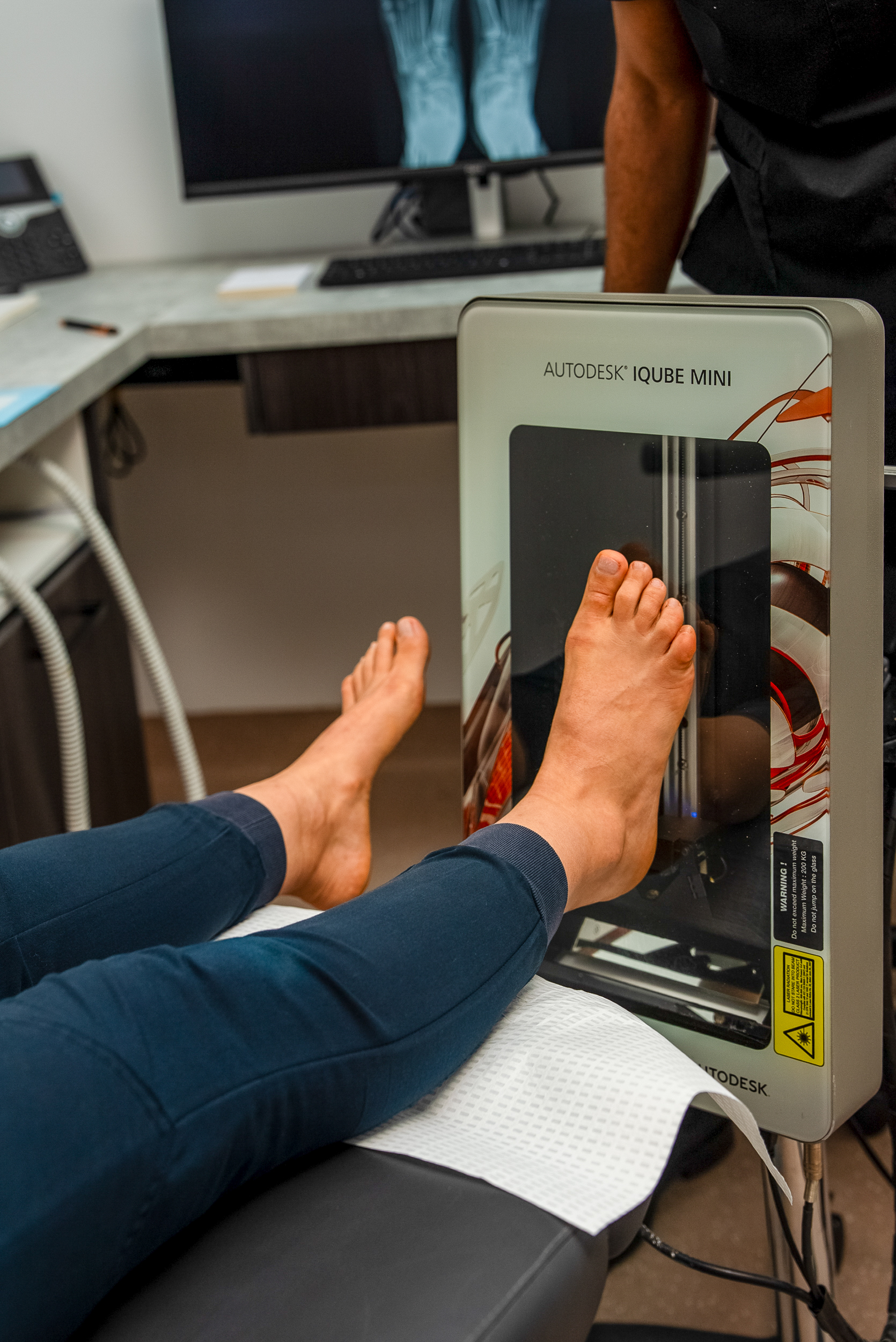
The confusion is reinforced by how X-rays work. An X-ray shows bone clearly but cannot image soft tissue. When a heel X-ray is taken, the spur appears prominently - a visible, defined, easily-labelled structure. The inflamed and degenerated plantar fascia is completely invisible. So the radiological report mentions the spur, the spur gets the diagnosis, and the actual cause of the pain goes unaddressed.
This misattribution has real consequences for management. Patients spend months trying to treat a structure (the spur) that isn't the source of their pain, while the actual problem (the fasciopathy) continues to worsen.
Is the Heel Spur Causing the Pain?
In most cases, no. The research on this is clear:
- Studies have found calcaneal spurs in approximately 11-16% of the asymptomatic adult population - people with no heel pain whatsoever. The spur being present does not predict pain.
- Conversely, plantar fasciitis is well-documented in people with no radiological evidence of a heel spur at all.
- Prospective studies tracking heel spur development and pain over time have found no consistent correlation between spur size and pain severity.
- Surgical removal of heel spurs without addressing the fascial pathology rarely produces lasting relief - further evidence that the spur was not the pain generator.
The American Orthopaedic Foot and Ankle Society and the Australian Podiatry Association both reflect current consensus: the heel spur is almost always incidental, and treatment should target the plantar fascia.
Are There Cases Where the Spur Itself Is Causing Pain?
Yes, in some specific circumstances. Insertional Achilles tendinopathy - a different condition from plantar fasciitis - involves spur formation at the posterior calcaneus (back of the heel), and in some cases the spur does directly contribute to pain by impinging on the overlying tendon. This is different from the plantar (inferior) spur associated with plantar fasciitis.
Additionally, in rare cases a very large plantar spur can cause direct soft tissue irritation or bursa formation adjacent to it. But these are the exception, not the rule, and even then surgery is considered only after exhaustive conservative management has failed.
Diagnosing the Real Cause of Your Heel Pain
A clinical diagnosis of plantar fasciitis does not require imaging. The diagnosis is made on the basis of:
- Location of pain - medial plantar heel, at the fascial origin
- Pattern of pain - worst with first steps in the morning, eases with movement, returns after prolonged standing
- Provocation on palpation - focal tenderness at the calcaneal attachment
- Absence of features suggesting alternative diagnoses (nerve entrapment, stress fracture, systemic arthritis)
X-ray is not required for the initial diagnosis of plantar fasciitis. It becomes useful when the diagnosis is uncertain, when symptoms are not responding as expected to appropriate treatment, or when a stress fracture needs to be excluded. If a spur is found, it should be documented but not automatically blamed.
Treatment - What Actually Works
Treatment is directed at the plantar fascia, not the spur. The evidence-supported approach for plantar fasciitis includes:
- Load management: Reducing the provocative activity to allow the tissue to settle without complete rest
- Stretching programme: Calf and plantar fascia stretching, particularly a morning pre-loading stretch, significantly reduces symptom severity
- Custom foot orthotics: Prescribed to offload the fascial origin and address the biomechanical drivers of the condition. See our detailed post on what custom orthotics are.
- Extracorporeal shockwave therapy (ESWT): The highest-evidence intervention for chronic plantar fasciitis. 70-80% response rates in clinical trials for presentations that have failed conservative management.
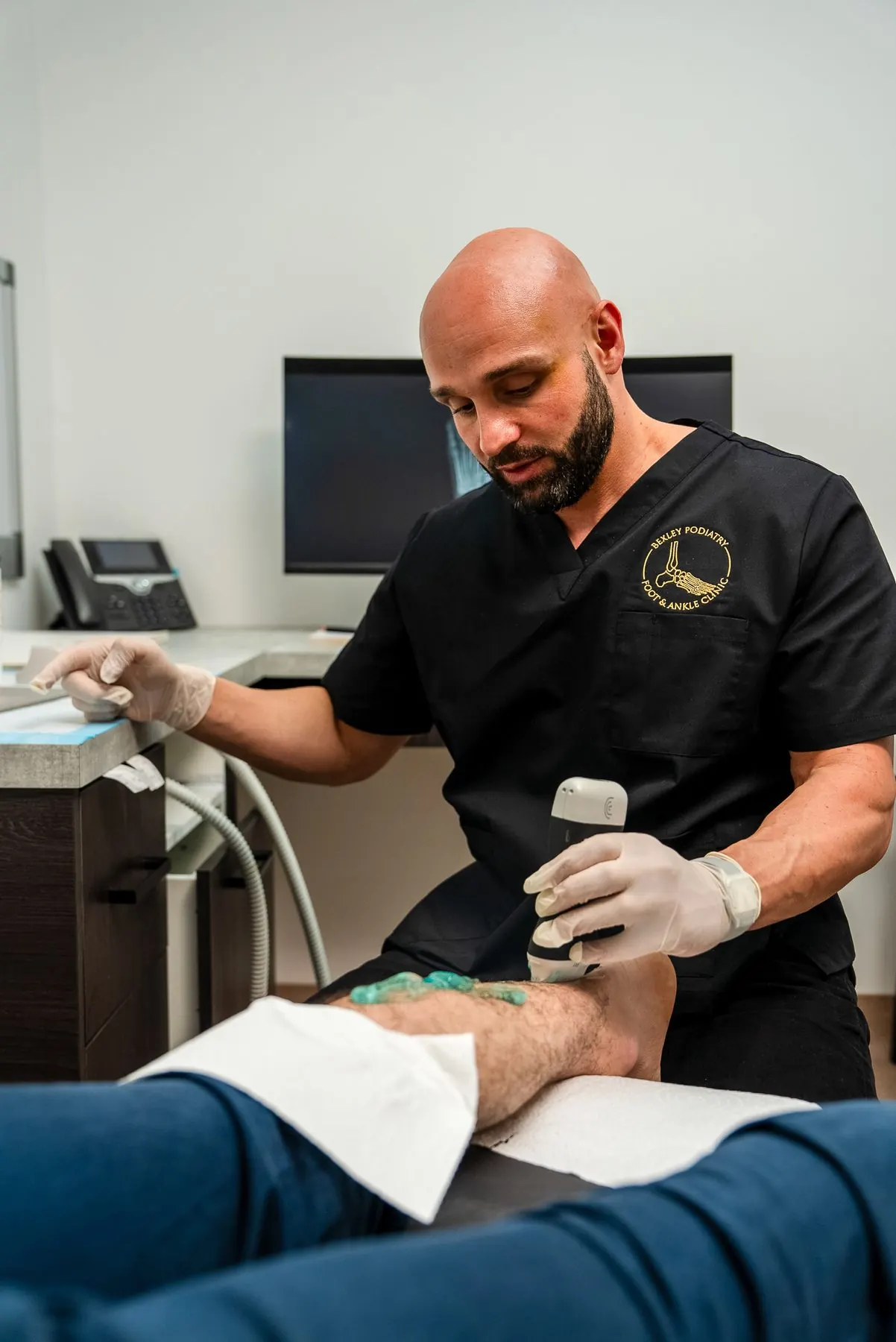
- Ultrasound-guided cortisone injection: Appropriate for acute, severe presentations to rapidly reduce symptom intensity so rehabilitation can proceed. Not a standalone cure.
- Surgical plantar fascia release: Reserved for genuinely refractory cases after 6-12 months of structured conservative management. Involves sectioning part of the fascial origin - not removing the spur. Success rates for well-selected patients are approximately 70-85%.
Stop treating the spur. Start treating the actual problem. A proper clinical assessment tells you exactly what's driving your heel pain and what intervention gives you the best chance of a lasting fix. Get a proper heel diagnosis.
Related on our site
Frequently asked questions
Should I have my heel spur removed?
Almost certainly not. The research does not support routine heel spur excision for plantar heel pain. The spur is not the primary pain generator in most cases. Treating the plantar fascia effectively - with load management, stretching, orthotics and if needed shockwave therapy - resolves symptoms for the vast majority of patients without surgery.
Will a heel spur keep growing?
Heel spurs grow slowly in response to sustained fascial tension. Once the underlying biomechanical loading is addressed through treatment, spur formation typically stabilises. Spurs do not usually continue growing once the provocative stress is removed.
How long does it take to recover from plantar fasciitis?
With appropriate early treatment: 6-12 weeks for most acute presentations. Chronic cases lasting 6 months or more may take 3-6 months of structured management. Shockwave therapy significantly accelerates recovery in persistent cases that have failed conventional approaches.
Can I have both plantar fasciitis and Achilles tendinopathy at the same time?
Yes. The two conditions share risk factors - particularly tight calf muscles and reduced ankle dorsiflexion - and can present simultaneously. They are separate diagnoses with overlapping management strategies. A clinical assessment will differentiate them and construct a treatment plan that addresses both.
Does cortisone injection cure plantar fasciitis?
Not in isolation. Cortisone injection provides rapid short-term pain reduction by suppressing the local inflammatory component, but does not address the underlying fascial degeneration. Evidence consistently shows that cortisone injections without concurrent rehabilitation produce high recurrence rates. They're best used to bring acute symptoms under control so exercise rehabilitation can begin.